Acute Patella Dislocation
What is the normal structure and function of a knee joint?
The patella (knee cap) sits within a groove in the front of the knee within the quadriceps tendon. As the knee moves the patella moves up and down in this groove. This movement is called “patella tracking”. The muscle that control this movement is the quadriceps muscle.
The quadriceps muscle is the large muscle at the front of the thigh. It is made up of four parts. These parts attach to the patella and form one common tendon that attaches to the shin. Contraction of the quadriceps muscle straightens the knee and thus moves the patella within the patella groove.
What is an Acute Patella Dislocation?
An acute patella dislocation occurs when the patella moves sideways out of its patella groove. It usually occurs with a twisting injury or direct blow.
Who gets it?
An acute patella dislocation can occur evenly between males and females, and occurs most often in teenagers. Patients who experience a dislocation are much more likely to develop chronic instability.
How does the injury present?
A dislocated patella is usually painful and causes swelling and deformity of the knee. Often the patella will move back into place (reduce) spontaneously. However, sometimes your child will require care from an Emergency Department at a hospital to manage the patella dislocation.

The appearance of an acutely dislocated patellar
What tests will need to be done?
X-rays will be taken initially to exclude the presence of a bony fragment or fracture and that the patellar is back in place.
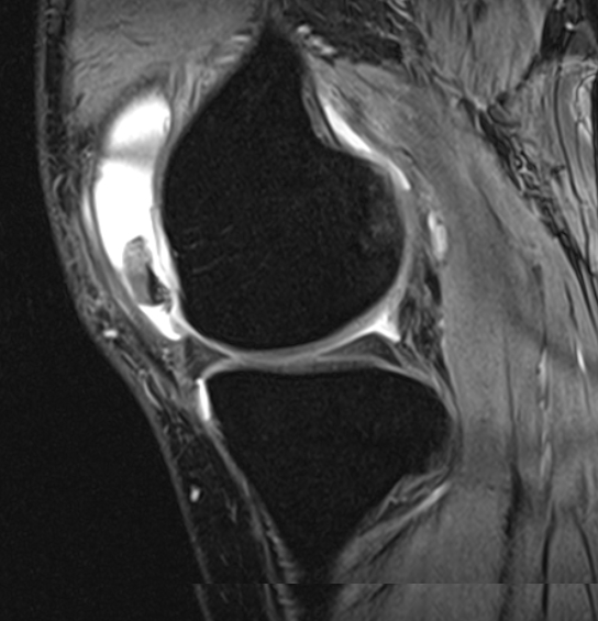
MRI (Magnetic Resonance Imaging) at the knee is also used for all first time dislocations within the first week from the injury. This is to assess there is any damage to the bone or cartilage. Damage to the bone or cartilage can occur as the patella moves out and back into its patella groove and affects up to 25 percent of children who have an acute patella dislocation.
MRI also provides information about the underlying structure of the patella and knee and provides some prognostic information about the likelihood of recurrent patella dislocation.
An MRI scan showing a large loose fragment of cartilage and bone
What is the treatment for a dislocated patella?
Once the patella is reduced, the knee is rested in a long leg splint and crutches are used to support the affected leg when walking for the first seven days. A regime of RICE (Rest, Ice, Compression and Elevation) for the initial 24-48 hours will assist in reducing pain and swelling of the knee. Physiotherapy should commence once the pain has eased.
An arthroscopy may be required if a loose bony fragment is identified which may require removal or refixation.
What will Physiotherapy involve?
Physiotherapy focusses on regaining range of motion and strengthening the inside part of the quadriceps muscle called the VMO. The VMO is weakened and the inside structures of the knee are damaged following an acute patella dislocation. This can cause the patella to track laterally (toward the outside) of the patella groove. Ongoing weakness of the VMO may put your child at risk of further patella dislocations.
When can the child return to playing sport?
Once your child is pain free and has regained normal quadriceps strength they can return to playing sport. This often takes approximately 8-12 weeks.
Children who experience ongoing patella dislocations may need Orthopaedic Surgical assessment and management.