 How is it classified?
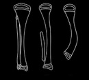
Achterman & Kalamachi classified fibular hemimelia into three radiological types depending of the deficiency of the fibular which was associated with increase severity of both the deformity and shortening of the limb
Unfortunately this system did not place importance on the foot nor did it correlate to then treatment strategy required to address the problem
How do you assess?
Careful clinical examination of the baby that is repeated over time usually gives the important details that allows initial treatment plans to be made. We will usually be able to inform parents on how many surgeries and of what type will, be required during childhood.
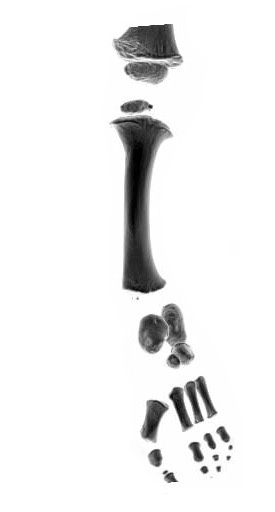
The initial focus is on the foot to assess its underlying deformity in what is a complex 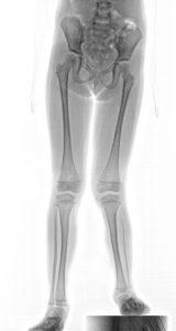 set of joints controlled by multiple tendons. To do this plain radiographs. CT scans and MRI are required but not usually until the child is about three years of age when surgery would often be performed.
Assessment of leg length at this age will give an indication of final leg length difference. Limb lengthening is usually the most reliable part of the treatment strategy.
The most essential part of assessment is to determine that a “good” foot can be achieved that will remain robust into adult life.
Children will function remarkably well when they are light and fit even with the most severe forms of fibular hemimelia. A small or unbalanced foot may not stand up to the rigours of adolescent and adult life therefor the importance of proper assessment and consultation with a clinician who is very familiar with the condition cannot be overemphasised
How do you treat fibular hemimelia?
Treatment strategies vary 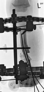 according to the type of fibular hemimelia. For some children it will be simply managing a leg length discrepancy with a shin lengthening procedure for others complex foot reconstruction, ligamentous stabilisation and repeated lengthening are required.
Some children with very poor feet are better off managed prosthetically but with advanced techniques now available these are much rarer event.
The majority of children with fibular hemimelia are suitable for reconstructive surgery and can expect to maintain very functional limbs of equal length into adulthood.